Pathologically altered tissues differ not only in morphology, including their shape and
the intensity of ultrasound reflections (echogenicity). In most cases, their consistency
(stiffness and elasticity) also changes. Most importantly, neoplastic lesions –
especially malignant tumors – usually show the highest tissue stiffness.
What is elastography?
Elastography is a diagnostic method that provides information about tissue stiffness and overlays this information onto morphological images, most commonly in the form of a transparent color map.
Tissue stiffness can be assessed using two main approaches:
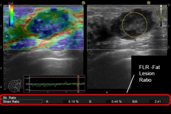
- Observation of tissue deformation in response to external mechanical forces (Strain Elastography).
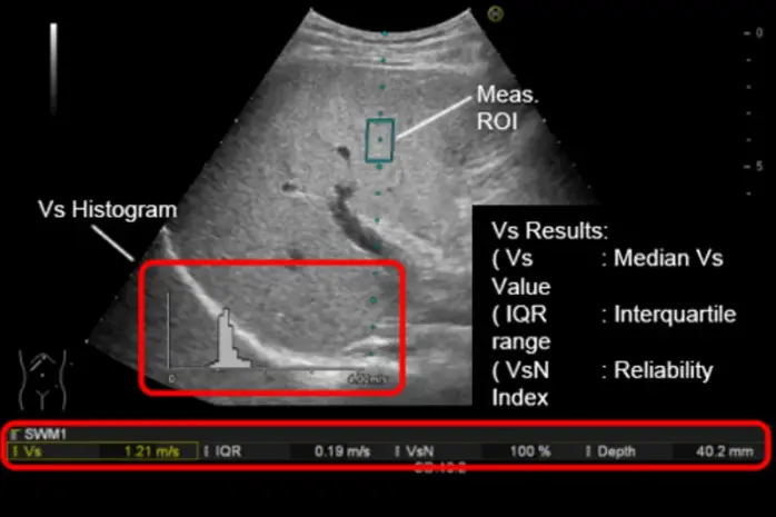
- Measurement of the propagation speed of waves within tissue, generated by a
high-frequency impulse (e.g., Shear Wave Elastography) or through Transient Elastography, commonly used for liver stiffness assessment.
When is strain elastography used?
Strain elastography is particularly useful when examining structures located close to the surface of the ultrasound probe.
This includes not only superficial organs such as:
- breasts
- thyroid gland and salivary glands
- testes and epididymis
- superficial lymph nodes
- elements of the musculoskeletal system
- skin and subcutaneous tissues
but also structures examined with probes inserted into body cavities close to the
organs of interest, for example:
- female reproductive organs
- prostate gland
- rectal wall in transrectal examinations
- walls of the esophagus, stomach, and duodenum, as well as upper abdominal
structures during endoscopic ultrasound (EUS).
In upcoming articles, we will discuss the application of elastography in specific
diagnostic situations.